
中

Recently, A team of scientists from the University of the Andes in Chile published a study entitled "Clinical‑grade extracellular vesicles derived from umbilical cord "in the Journal of Nanobiotechnology mesenchymal stromal cells: preclinical development and first‑in‑human intra‑articular validation as therapeutics for knee Study of osteoarthritis (Umbilical cord mesenchymal stromal cell clinical-grade exosomes: preclinical development and first human intraarticular validation as a treatment for knee osteoarthritis).
In this study, the research team conducted a comprehensive investigation of umbilical cord mesenchymal stromal cell (UC-MSC) -derived exosomes (SEVs) with the help of a variety of in vitro and in vivo experiments. The research content covers its efficacy and safety in the treatment of knee osteoarthritis, as well as digging into the potential mechanism of its role, and is committed to opening up a new path for the treatment of knee osteoarthritis.
01 Osteoarthritis
Osteoarthritis (OA), as a common and progressive multi-factor joint disease, is one of the key factors that cause chronic pain and lead to disability. Among the many affected joints, the knee is the most severely affected part of OA, and nearly 4/5 of the world's OA cases occur in the knee. At present, there is no treatment for knee osteoarthritis that can delay cartilage degradation or restore joint cartilage function. Existing treatment options rely primarily on a multimodal approach aimed at controlling pain and relieving symptoms of joint stiffness.
The salient features of OA include chronic synovitis and cartilage degeneration, and there is growing evidence that synovitis precedes cartilage degeneration. Macrophages play a key role in synovial tissue. Cytokines such as IL-1β, IL-6 and TNF-α released by proinflammatory M1 subtype macrophages can trigger, maintain and amplify inflammation. IL-10 secreted by M2 macrophages with an anti-inflammatory phenotype is essential for tissue repair and inflammation resolution. In OA patients, synovitis is manifested by an increase in the number of M1 macrophages that produce reactive oxygen species (ROS), which can lead to inflammation, stromal dysregulation and cartilage injury, and oxidative stress can induce apoptosis of primary chondrocytes. Therefore, controlling inflammation and oxidative stress has become a key strategy for OA treatment.
Mesenchymal stromal cells (MSC) and the sEV they secrete have shown potential in OA therapy. MSC-sEV has tissue regeneration capabilities similar to cells and is safer as a cell-free treatment option. In animal models of OA, MSC-sEV therapy has been shown to promote cartilage regeneration and slow the progression of OA. Umbilical cord (UC) derived MSCS are considered to be the most potential source of MSCS for the treatment of OA due to their strong chondrogenic ability, in vitro inhibition of T cell proliferation, and relatively low angiogenic properties. Based on this, the research team aims to develop a clinical-grade therapy based on UC-MSC-derived sEV and evaluate its efficacy and safety.
02 Research Data
In this research exploration, the research team aims to develop a clinical-grade therapy based on UC-MSC-derived sEV, and further explore its efficacy and safety.
To this end, the researchers conducted a comprehensive characterization of UC-MSC-derived sEV in non-CGMP conditions. The study found that multiple batches of sEV showed consistent miRNA and protein profiles, a key achievement that laid a solid foundation for subsequent standardized production processes and greatly improved the feasibility of UC-MSC-derived sEV therapies towards clinical application.
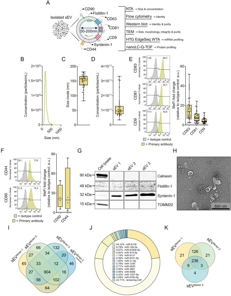
UC-MSC secretes sEV with a unique and replicable molecular cargo
In vitro experiment
In vitro experiments, sEV demonstrated chondroprotective activity. The research team established a polarized human monocyte derived macrophage (hmMΦs) model. Flow analysis showed that the expression of M1 pro-inflammatory markers (CD86, HLA-DR) decreased and M2 anti-inflammatory markers (CD206, CD163) increased in the hmMΦs treated with sEV. ELISA assay showed that the secretion of anti-inflammatory factor IL-10 increased, and the proinflammatory factors IL-6, TNF-α and IL-1β decreased.
In addition, in menadione-induced cytotoxicity tests, sEV can effectively protect chondrocytes, resulting in a significant reduction in the proportion of apoptotic/dead cells. Moreover, the cytotoxicity test results based on LDH release showed that sEV did not show significant toxicity to chondrocytes.
The results of this series of in vitro experiments fully confirmed that sEV has a positive and effective role in cartilage protection.
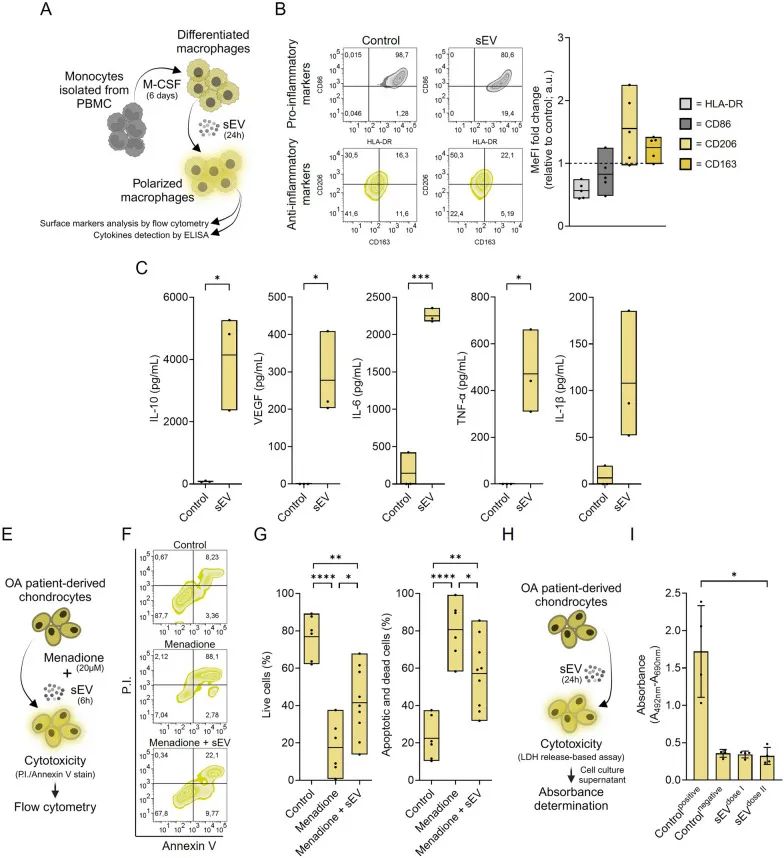
sEV drives the polarization of macrophages and exerts chondroprotective activity against oxidative stress
Mouse experiment
In the mouse experimental phase, the research team constructed a mouse model of collagenase-induced osteoarthritis (CIOA) and conducted preclinical experiments with intra-articular administration of sEV. Using μCT image analysis, knee bone mineral density (BMD) and bone surface area to bone volume ratio (BS/BV) index were improved in sEV treated mice. At the same time, Safranin O/Fast green staining strongly confirmed the protective effect of sEV on the cartilage, which was associated with a lower histological score.
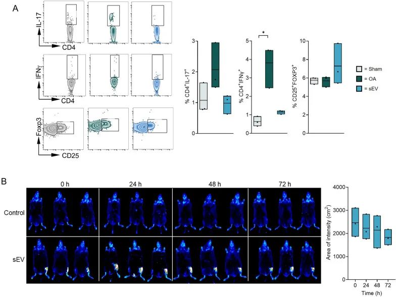
In addition, the results of the examination of the mouse posterior knee lymph nodes showed that sEV has an inflammatory effect and can effectively regulate the CD4+T cell population, which is manifested by reducing the number of pro-inflammatory cells and increasing the proportion of regulatory T cells (CD25+FOXP3+Treg cells). The results of biological distribution showed that sEV could be stable in the knee space of mice after injection of DIR-stained sEV.
These experimental results fully indicate that sEV plays a positive role in improving the symptoms of osteoarthritis in mice, protecting cartilage and regulating immune inflammatory response, which provides an important experimental basis for its subsequent clinical application in the treatment of osteoarthritis.
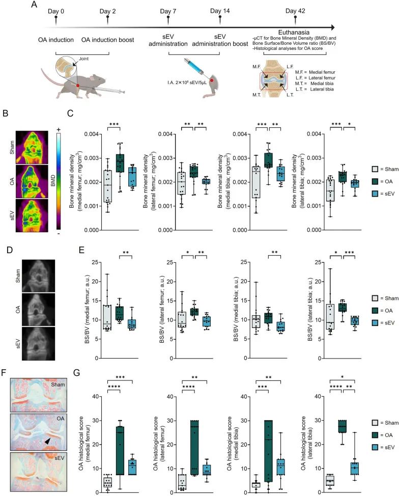
sEV reduces the severity of osteoarthritis and promotes osteoarthritis regeneration in an in vivo mouse model
Human clinical trial
In the first human clinical trial, the research team injected clinically-grade UC-MSC-sEV into the joints of patients with knee osteoarthritis. The results showed that patients' clinical score index (WOMAC index) decreased and symptoms improved before administration and after 1 year of treatment, and no adverse reactions were observed. The third-party MRI assessment of the SPAIR and WATSc sequences found no signs of cartilage degradation in the patient, demonstrating the initial safety of the first human intraarticular administration of sEV.
Based on this, the research team also designed a further phase 1 clinical trial. In the follow-up trial plan, patients will receive three different doses of sEV (2±0.5E + 9 particles/3 mL, 6±0.5E + 9 particles/3 mL, 20±0.5E + 9 particles/3 mL), respectively. The patients were followed up for 1 year. It is worth mentioning that these preliminary trial results were further validated in the Phase 1/2 randomized controlled clinical trial and the Phase 1 dose-escalation clinical trial conducted in 2019.
1. Study on sEV uptake
In order to further explore the uptake mechanism of sEV, the research team used sEV tracer reagents to label it, and then observed the dynamic process of sEV entering osteoarthritis related cell lines. When mirNA-loaded sEV entered the cell, miRNA was detected by real-time fluorescence quantitative polymerase chain reaction (qPCR). The experimental results confirm that sEV can not only enter the cell smoothly, but also carry the cargo with biological function, which lays the foundation for its therapeutic effect.
2. Studies on macrophage polarization and chondrocyte protection
During the study, it was found that MSC-sEV has a unique function of driving the polarization of macrophages. It was able to significantly increase the proportion of M2-polarized macrophages with anti-inflammatory activity, while prompting M2 macrophages to secrete more interleukin-10 (IL-10). IL-10 has a positive effect on tissue repair and inflammation regression, and then effectively protects chondrocytes in oxidative stress environment, providing a new way for the treatment of OA.
3. Transcriptomic analysis
Using transcriptomics, the research team analyzed the enrichment patterns of 16 common miRNAs in MSC-sEV RNA in depth and performed gene ontology (GO) analysis of the proteins they regulate. Through this study, multiple biological processes and presumed target genes closely related to macrophages and inflammatory processes were successfully revealed, providing key clues for further understanding of the mechanism of action of MSC-sEV.
4. Animal model experiment
To verify the therapeutic effect of MSC-sEV in vivo, the research team built a mouse model of arthritis and intervened by giving MSC-sEV in the joint. During the experiment, micro-computed tomography (μCT) was used to accurately analyze the bone density of the joint area to evaluate joint regeneration and cartilage repair. The animal results provide strong in vivo evidence for the potential application of MSC-sEV in the treatment of OA.
5. Study on immunosuppressive activity and stability
In order to explore the immunosuppressive activity and stability of MSC-sEV, the research team injected MSC-sEV into the joint cavity of mice, and then measured the levels of CD4+IL-17+ cells and CD4+IFN-γ+ cells to evaluate its immunosuppressive activity. At the same time, sEV tracer analysis was used to determine the long-term stability of sEV in the joint cavity. The results showed that MSC-sEV could persist in joint lumen for a long time and exert immunosuppressive effect continuously.
6. Verify the storage stability
Considering the needs of MSC-sEV for future commercialization and clinical applications, the research team conducted storage stability verification. Through a series of experiments, the quality and activity of MSC-sEV can be effectively maintained in the storage process, which provides an important guarantee for its subsequent practical application.
7. Design of the first human phase 1 clinical study
In order to advance MSC-sEV to clinical application, the research team carefully designed the first phase 1 clinical study of MSC-sEV administration in humans. In the study design, the different dose groups were clearly divided and the follow-up time was determined, which provided a scientific and rigorous basis for the follow-up large-scale clinical trials, and promoted the transformation process of MSC-sEV from laboratory research to clinical treatment.
sEV has immunosuppressive activity in vivo and is maintained within the knee space
The study comprehensively evaluated the efficacy, safety and potential mechanisms of UC-MSC-derived sEV for the treatment of knee osteoarthritis through a variety of in vitro and in vivo trials. For the first time, the safety of a clinical-grade product for intra-articular administration in humans was evaluated, and the complete characterization of UC-MSC-derived sEV in non-CGMP conditions was established, as well as the feasibility of a standardized production process.
Studies have fully confirmed the regenerative and anti-inflammatory properties of UC-MSC-sEV as an intra-articular administration therapy, as well as a good safety profile. The follow-up phase 1 clinical study of different doses of sEV treatment will further explore the best scheme for the treatment of knee osteoarthritis, laying a solid foundation for the future wide application of sEV in the treatment of knee osteoarthritis.